
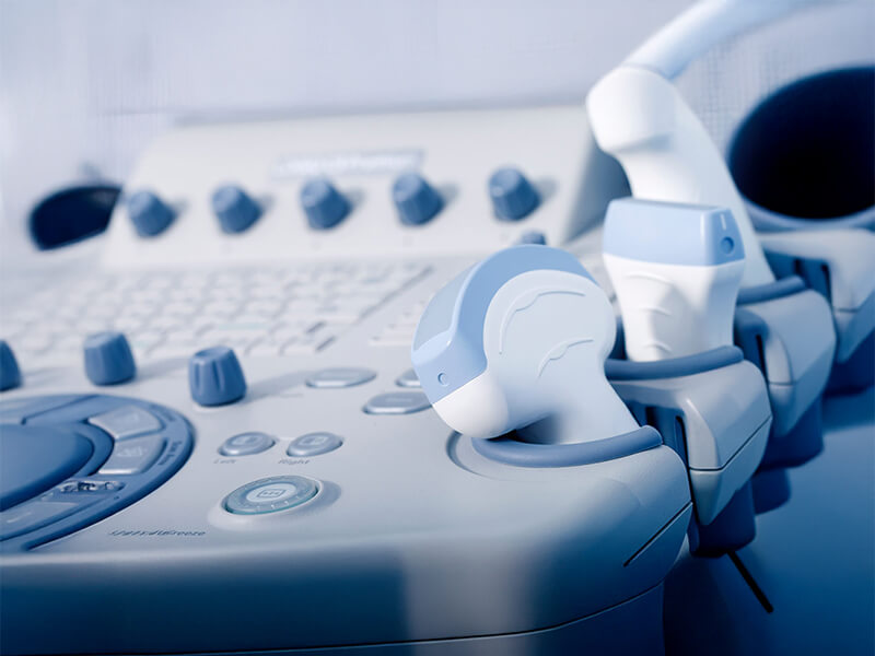
Ultrasound Scans (Sonograms)

An ultrasound scan is a fundamental, non-invasive diagnostic method. It allows for precise assessment of the shape, size, and depth of individual organs. Ultrasound scans are also used in prenatal examinations due to their complete safety for pregnant women and lack of impact on the foetus. Thanks to modern equipment, a reliable three-dimensional (3D or 4D) image of the organ being examined can be obtained. Ultrasound scans can be repeated multiple times for the same individual, enabling doctors to detect pathological changes in internal organs at an early stage. 

Diagnostic tests can also be performed preventively to provide peace of mind, even when no symptoms are present.

Types of Ultrasound Scans:

Paediatric Ultrasound Scans

A cranial suture ultrasound is a completely safe and painless examination that allows for the assessment of an infant’s head. The ultrasound clearly visualises the connections between the skull bones, known as cranial sutures. An abnormal head shape observed by a paediatrician or parent may result from a defect in the sutures, such as their premature fusion. The ultrasound enables early diagnosis and treatment before a significant skull deformation occurs. If the cranial sutures are normal (the head deformity is not due to a suture defect), treatment for the infant’s head curvature can be undertaken using helmet therapy. This is a modern method of correcting head shape using a custom-made orthosis or helmet. Additionally, a cranial suture ultrasound can also visualise and verify lump-like changes, cysts, or atypically located lymph nodes, allowing for appropriate diagnostic and therapeutic measures. The ultrasound itself requires no preparation and can be performed simultaneously with a transcranial brain ultrasound. 

A cranial ultrasound, also known as transfontanellar ultrasound, is an examination performed on new-borns and infants to assess the anatomy and development of their brains. This examination is most commonly performed on children born prematurely, those who experienced birth trauma, or those diagnosed with neurological disorders. Indications for this examination also include maternal gestational diabetes, placental abruption, or intrauterine infections during pregnancy. 

A hip ultrasound is a recommended examination for all infants due to the frequent occurrence of abnormalities (approximately 1 in 200 children). This examination should be performed no later than 6 weeks of age. During the examination, the anatomical structures and maturity of the hip joint are assessed. It is particularly important to perform this examination in infants with a limited range of motion in the hip joints, differences in the structure or length of the lower limbs, or infants exhibit a clicking sound during movement.

An abdominal ultrasound is currently the most common type of ultrasound performed in daily medical practice, as it allows for the visualisation of internal organs such as the liver, gallbladder and bile ducts, spleen, pancreas, kidneys, adrenal glands, prostate, bladder, and uterine adnexa. During the examination, large blood vessels (e.g., the aorta) and lymph nodes are also examined. The stomach and intestines can also be examined although only partially. The most common indications for this type of ultrasound in patients include: 

  • Abdominal pain 
  • Nausea and vomiting 
  • Palpable abnormalities during a physical examination of the abdomen 
  • Abdominal trauma 
  • Increased abdominal circumference 
  • Unexplained weight loss 
  • Difficulty urinating or passing stools
  • Bleeding from the gastrointestinal tract, urinary system, or reproductive organs
  • Abnormal blood test results 
  • Chronic conditions – monitoring
  • Further diagnostics for extra-abdominal conditions 

Preparation for an abdominal ultrasound is essential: adults and teenagers should fast for 6 hours before the examination and reduce gases in the digestive system using over-the-counter oral medications. A few days before the examination, patients should follow a light diet. Drinking still water to fill the bladder is also recommended, as this is necessary for bladder, prostate, or ovaries imaging. Younger children, especially under 3 years of age, require no preparation.

This examination assesses the inguinal canals and scrotum, often used to diagnose hernias, undescended testicles, or other abnormalities in the groin area. 

This examination assesses the thyroid gland, salivary glands, and lymph nodes, helping to identify abnormalities such as nodules, inflammation, or tumours. 

A pelvic ultrasound is used to assess the organs within the pelvis, including the bladder, uterus, ovaries, and prostate. It is commonly used to diagnose conditions such as cysts, fibroids, or abnormalities in the reproductive or urinary systems.

Gynaecological Ultrasound Scans

A pelvic ultrasound used to assess the bladder, uterus, ovaries, and adnexa. It is commonly used to diagnose cysts, fibroids, or abnormalities in the reproductive or urinary systems.

A breast ultrasound should be performed regularly, once a year, for all women over the age of 35. Currently, it is the primary diagnostic examination for the early detection of breast cancer. The examination allows for the assessment of the structure of glandular and fatty breast tissue, milk ducts, and lymph nodes. It can be performed on any day of the menstrual cycle, but for preventive screenings, it is best to schedule it between the 1st and 10th day of the menstrual cycle. This is a completely painless and safe examination for patients, enabling the detection of even the smallest changes in the breasts that are not palpable during a physical examination. Absolute indications for a breast ultrasound include: breast pain, a lump or other changes in the breast, changes in the skin of the breast, or discharge from the nipple.

Evaluation of nipple changes or discharge.

Ultrasound tracking of ovulation and menstrual health.

Safe prenatal exam, high-quality 3D/4D foetal imaging.

Cardiac Ultrasound Scans

Assessment of vascular structures in upper limbs.

Assessment of vascular structures in lower limbs.

This is an ultrasound examination of the heart’s structures and movements. It evaluates the heart’s anatomy, valve function, and the contractility of the heart muscle. It is particularly recommended for patients suspected of having heart disease.

Endocrinological Ultrasound Scans

A thyroid ultrasound is a diagnostic examination that enables the doctor to assess the size and structure of the thyroid gland. Additionally, it allows for the visualisation of any abnormalities within the gland, such as nodules or cysts. Doppler imaging is always used during a thyroid ultrasound to evaluate blood flow and the degree of vascularisation of the gland.
If focal changes in the thyroid, such as nodules, are detected, the patient may be referred for a fine-needle aspiration biopsy to assess the cellular composition of the lesion through microscopic (cytological) examination. The biopsy is always performed under ultrasound guidance, ensuring precision and safety. Cytological evaluation of a thyroid lesion helps to rule out malignancy or confirm suspicions, allowing for appropriate further management and treatment.
The most common indications for a thyroid ultrasound include abnormal thyroid hormone or TSH levels, the presence of a neck lump, hoarseness, an enlarged neck contour, restricted neck movement, neck pain, or discomfort when swallowing or breathing.

Imaging of parathyroid glands

Imaging of adrenal gland structure.

Assessment of salivary glands.

Evaluation of thyroid enlargement and nodularity.

Nephrological & Urological Ultrasound Scans

  • Kidney Ultrasound
  • Prostate Ultrasound
  • Urethral Ultrasound
  • Inguinal Canals and Scrotum Ultrasound
  • Urinary Tract Ultrasound
  • Testicular Ultrasound
  • Scrotal Ultrasound

Orthopaedic Ultrasound Scans

  • Joint Ultrasound
  • Tendon Ultrasound
  • Muscle Ultrasound
  • Ligament Ultrasound

Surgical Ultrasound Scans

A thyroid ultrasound is a diagnostic examination that enables the doctor to assess the size and structure of the thyroid gland. Additionally, it allows for the visualisation of any abnormalities within the gland, such as nodules or cysts. Doppler imaging is always used during a thyroid ultrasound to evaluate blood flow and the degree of vascularisation of the gland.
If focal changes in the thyroid, such as nodules, are detected, the patient may be referred for a fine-needle aspiration biopsy to assess the cellular composition of the lesion through microscopic (cytological) examination. The biopsy is always performed under ultrasound guidance, ensuring precision and safety. Cytological evaluation of a thyroid lesion helps to rule out malignancy or confirm suspicions, allowing for appropriate further management and treatment.
The most common indications for a thyroid ultrasound include abnormal thyroid hormone or TSH levels, the presence of a neck lump, hoarseness, an enlarged neck contour, restricted neck movement, neck pain, or discomfort when swallowing or breathing.

Non-invasive vascular ultrasound of the neck.

Assessment of superficial nodules and soft-tissue lesions.

Evaluation of lymph node size and morphology.

An abdominal ultrasound is currently the most common type of ultrasound performed in daily medical practice, as it allows for the visualisation of internal organs such as the liver, gallbladder and bile ducts, spleen, pancreas, kidneys, adrenal glands, prostate, bladder, and uterine adnexa. During the examination, large blood vessels (e.g., the aorta) and lymph nodes are also examined. The stomach and intestines can also be examined although only partially. The most common indications for this type of ultrasound in patients include: 

  • Abdominal pain 
  • Nausea and vomiting 
  • Palpable abnormalities during a physical examination of the abdomen 
  • Abdominal trauma 
  • Increased abdominal circumference 
  • Unexplained weight loss 
  • Difficulty urinating or passing stools
  • Bleeding from the gastrointestinal tract, urinary system, or reproductive organs
  • Abnormal blood test results 
  • Chronic conditions – monitoring
  • Further diagnostics for extra-abdominal conditions 

Preparation for an abdominal ultrasound is essential: adults and teenagers should fast for 6 hours before the examination and reduce gases in the digestive system using over-the-counter oral medications. A few days before the examination, patients should follow a light diet. Drinking still water to fill the bladder is also recommended, as this is necessary for bladder, prostate, or ovaries imaging. Younger children, especially under 3 years of age, require no preparation.

Contrast-Enhanced Ultrasound (CEUS)

A Contrast-Enhanced Ultrasound (CEUS) is a specialised imaging technique that uses a contrast agent to improve the visibility of blood flow and tissue structures.

How to Prepare for Ultrasound Scans

  • The examination should be performed on an empty stomach (the last meal should be eaten no later than 5 hours before the ultrasound). 
  • Avoid chewing gum or smoking cigarettes before the examination. 
  • Palpable abnormalities during a physical examination of the abdomen 
  • Abdominal trauma 
  • Increased abdominal circumference Patients taking medications should take them as prescribed by their doctor, but no later than 1 hour before the examination. 
  • On the afternoon/evening before the examination, avoid foods and drinks that cause bloating, carbonated beverages, yoghurts, etc. 
  • The examination is performed with a full bladder, so approximately 1-1.5 litres of still water should be drank one hour before the examination. During this time, do not urinate. 
  • For a transrectal examination, the patient should empty their bowels beforehand (an enema is recommended). 
  • For an abdominal examination, no special preparation is required. 
  • It is best to perform the examination in the first half of the menstrual cycle (between the 5th and 10th day of the cycle). 
  • Patients taking medications should take them as prescribed by their doctor, but no later than 1 hour before the examination, with still water. 
  • For examinations of the renal artery, aorta, portal vein, or hip joints, the patient should fast for 6 hours before the examination. 

Disclaimer
*The Pax Medicus website provides information for guidance only and does not constitute medical advice, diagnosis, or treatment. Pax Medicus does not provide healthcare services and is not a substitute for consultation with a qualified medical professional. Always seek advice from your physician regarding any medical condition. Prices shown are indicative only and subject to confirmation by the healthcare provider. This is the official website of Pax Medicus; we do not operate through agents or third-party sites.

BOOK YOUR CONSULTATION TODAY


Simply fill in the form below and our team will get back to you as soon as possible.