


Intervertebral Disc Herniation Surgery
At Pax Medicus, we connect you with trusted specialists offering advanced treatments for intervertebral disc herniation. Our minimally invasive procedures aim to relieve pain, restore mobility, and prevent future complications, helping you return to your daily activities with confidence.
What is an Intervertebral Disc?
The spine consists of vertebrae (bones) and intervertebral discs, which are cartilage structures positioned between each vertebra. These discs allow movement between the vertebrae, enabling flexion and extension of the torso and neck. Additionally, they absorb some of the forces acting on the spine, reducing the pressure on the vertebrae.
Each intervertebral disc is made up of two main parts: the nucleus pulposus, a soft, elastic core, and the annulus fibrosus, which is the tough outer layer that holds the nucleus in place. The nucleus pulposus acts as a cushion due to its high water content, while the annulus fibrosus resists pressure and stretching forces.
What is a Herniated Disc?
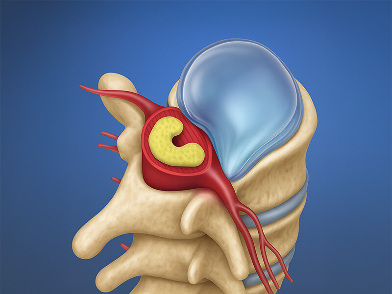
A herniated disc occurs when the nucleus pulposus protrudes or ruptures through the annulus fibrosus. Depending on the severity of the protrusion, a herniated disc can be classified into three stages:
What are the Symptoms of a Herniated Disc?
Symptoms of a herniated disc are mainly caused by compression of the nerve roots and spinal cord. The most common symptom reported by patients is pain, which can be localised in the spine or radiate to the arms or legs. In addition to pain, patients may also experience numbness or tingling sensations in the affected limb.
Pain often intensifies during physical activity, coughing, or sneezing due to increased intra-abdominal pressure, which further compresses the spine. Sitting may worsen the pain compared to standing, as the pressure on the spine is higher when seated
What are the Causes of a Herniated Disc?
A herniated disc typically develops due to repeated spinal stress over time. Factors such as obesity, frequent heavy lifting, and age-related degeneration of the disc make the annulus fibrosus more susceptible to damage, allowing the nucleus pulposus to protrude. Smoking accelerates the degenerative process of the discs, increasing the risk of injury.
How is a Herniated Disc Diagnosed?
To diagnose a herniated disc, imaging tests are required. The first step is an X-ray, which can rule out other potential issues like fractures or infections. Although an X-ray cannot directly visualise a herniated disc, it can help spot indirect signs such as a loss of the spine’s natural curvature.
The gold standard for diagnosing a herniated disc is magnetic resonance imaging (MRI). MRI provides detailed images of the soft tissues, allowing for accurate assessment of the herniation, its size, and its impact on surrounding structures like nerve roots.
What Does Treatment of a Herniated Disc Involve?
The first-line treatment for a herniated disc is conservative treatment. This includes rest, avoiding heavy lifting, and using pain-relieving medications such as analgesics and muscle relaxants. In certain cases, glucocorticoid injections may be used to reduce inflammation and relieve pressure on the affected nerve roots.
Physiotherapy plays a vital role in recovery. Strengthening the muscles around the spine can help stabilise the lumbar region and reduce the risk of future disc injuries. Additionally, weight reduction and proper lifting techniques are crucial in preventing additional strain on the spine.
What are the Indications for Surgical Treatment of a Herniated Disc?
Surgery is considered when conservative treatment fails. It is typically indicated in cases of lumbar radicular syndrome, sensory disturbances in the lower limbs, foot movement restrictions, calf dysfunction, progressive sexual dysfunctions, or bladder dysfunction.
What Does Surgical Treatment of a Herniated Disc Involve?
The choice of surgical method depends on the location of the herniation, the severity of symptoms, and the patient’s age and overall health.
Microdiscectomy is the most common surgical procedure used to treat lumbar disc herniation. It involves making a small incision (about 1–2 cm) above the affected area. The surgeon gently moves the paraspinal muscles aside and may create a small opening in the vertebra (laminotomy) or remove part of the bone (laminectomy) to access the damaged disc. The herniated disc fragment is then carefully removed to relieve pressure on the affected nerve root.
If degenerative changes have caused narrowing of the intervertebral foramen — the space through which nerves exit the spinal canal – a foraminotomy may be performed. This involves removing excess bone or tissue to restore proper nerve function. In cases where spinal stability is compromised, spinal fusion may also be required to strengthen and stabilise the spine.
Endoscopic discectomy is a less invasive alternative to traditional open surgery. It is performed through a small incision using an endoscope (a tiny camera) that provides real-time visuals on a monitor. Specialised surgical instruments are inserted through the same incision to remove the herniated disc material without the need for a laminotomy. This approach typically results in faster recovery and minimal tissue damage.
In some cases, artificial disc replacement may be considered – particularly for patients with a single-level disc herniation. The procedure is performed through a small abdominal incision (3–8 cm) without cutting through the abdominal muscles. The peritoneal sac, which holds the abdominal organs, is gently moved aside to access the front of the spine. The damaged disc is then removed, normal disc height is restored, and an artificial disc is implanted to preserve natural spinal movement.
Incision
1–2 cm (minimally invasive)
Procedure duration
Approximately 1 hour
Anaesthesia
General anaesthesia
Your journey step by step
History, examination & MRI review
Conservative care or surgery
Microdiscectomy or endoscopic discectomy
Early mobilisation & physiotherapy
Wound check, rehab & prevention
Wound check, rehab & prevention
What Is the Recovery Process After Microdiscectomy?
Microdiscectomy is a minimally invasive procedure with a relatively quick recovery time. Many patients are able to leave the hospital the same day or within 24 hours. During recovery, physiotherapists provide guidance on how to perform daily activities and exercises to promote healing and prevent strain on the spine.
Microdiscectomy is a minimally invasive procedure with a relatively quick recovery time. Many patients are able to leave the hospital the same day or within 24 hours. During recovery, physiotherapists provide guidance on how to perform daily activities and exercises to promote healing and prevent strain on the spine.
Disclaimer
*The Pax Medicus website provides information for guidance only and does not constitute medical advice, diagnosis, or treatment. Pax Medicus does not provide healthcare services and is not a substitute for consultation with a qualified medical professional. Always seek advice from your physician regarding any medical condition. Prices shown are indicative only and subject to confirmation by the healthcare provider. This is the official website of Pax Medicus; we do not operate through agents or third-party sites.
BOOK YOUR CONSULTATION TODAY
Simply fill in the form below and our team will get back to you as soon as possible.