
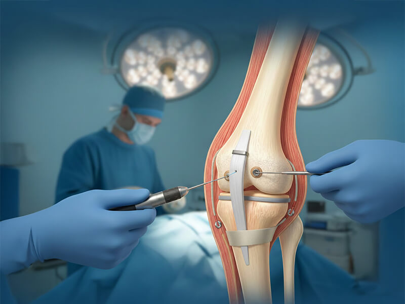
Surgical Treatment of Patellar Dislocation | PAX Medicus Medical Tourism

Surgical Treatment of Patellar Dislocation

Patellar dislocation occurs when the kneecap moves out of its normal position, causing pain, swelling and instability. Through our trusted partner hospitals, Pax Medicus coordinates modern stabilisation surgery and rehabilitation pathways abroad for patients with recurrent patellar dislocation.

As a medical tourism agency, we organise your diagnostics, surgical treatment and hospital stay, while your operation is carried out by experienced orthopaedic surgeons in accredited centres.

What Is Patellar Dislocation?

Patellar dislocation is when your kneecap slides out of its normal groove, usually to the outer side of the knee. It can happen after a sudden twist, a fall or a direct impact, and in some people it can become a repeated problem.

  • The kneecap can slip out of place and sometimes pop back by itself.
  • Typical symptoms include sudden pain, swelling and difficulty moving the knee.
  • The knee may feel unstable or as if it might “give way”.
  • Some patients develop repeated dislocations because of their anatomy.

Understanding the Patella (Kneecap)

Key facts:

  • Triangular bone at the front of the knee.
  • Largest sesamoid bone (formed inside a tendon).
  • Protects the knee joint.
  • Improves the strength of the quadriceps muscle.
  • Connects via the quadriceps tendon and patellar ligament to the shinbone.

Why Does Patellar Dislocation Happen?

A dislocation occurs when the kneecap moves out of its normal groove at the end of the femur. This is usually caused by a twisting injury.

Typical injury mechanism

  • Foot is firmly planted on the ground.
  • Thigh rotates inward.
  • Knee is bent and knock-kneed (valgus).
  • Quadriceps muscle contracts strongly.

Structures usually injured

  • Medial patellofemoral ligament (MPFL) – almost always damaged.
  • Cartilage on the kneecap.
  • Cartilage on the lateral femoral condyle.

Other causes & risk factors

  • Direct blow to the inner side of the knee in sport.
  • Long patellar ligament.
  • Knock-knee (valgus) alignment.
  • High-riding patella (patella alta).
  • Shallow femoral groove.

The TT–TG distance (tibial tuberosity–trochlear groove) helps assess alignment: values > 15 mm are abnormal, and > 20 mm are associated with recurrent instability.

Symptoms of Patellar Dislocation

Immediately after injury

  • Sudden sharp pain in the knee.
  • Feeling of the kneecap moving sideways.
  • Swelling due to bleeding in the joint.
  • Difficulty fully bending or straightening the knee.

Ongoing symptoms

  • Pain during walking, sport or long distances.
  • Discomfort when repeatedly bending the knee.
  • Sensation of “something moving” inside the joint.
  • Episodes of the knee giving way or feeling unstable.

A specialist will take a detailed history and examine the knee. Even if the kneecap has slipped back into place by itself, imaging is important to reveal hidden damage.

Clinical assessment

  • Examination of knee alignment.
  • Stability and range of movement tests.
  • Assessment of swelling and tenderness.

Imaging

  • X-rays (AP, lateral and Merchant views).
  • Ultrasound to assess effusion and soft tissues.
  • MRI if cartilage damage or ligament tears are suspected.

Yes, first-time patellar dislocation without fractures or loose cartilage fragments can often be treated conservatively.

Conservative treatment

  • Cold compresses to reduce pain and swelling.
  • Temporary reduction of activity and weight-bearing.
  • Structured physiotherapy to strengthen muscles.
  • Short-term use of a knee brace (while exercising regularly).

When it works best

  • First dislocation episode.
  • No major cartilage or bone damage.
  • Patient carefully follows medical advice.

Immediately after injury

  • Recurrent patellar dislocations.
  • Chondral or osteochondral fractures with loose fragments.
  • Significant anatomical abnormalities causing instability.
  • Significant anatomical abnormalities causing instability.

The overall goal of surgery is to restore patellar stability, correct abnormal alignment and protect the cartilage from further damage.

Surgical Options for Patellar Instability

1. Arthroscopic lateral release

  • Minimally invasive keyhole surgery.
  • Releases the tight lateral retinaculum.
  • Reduces excessive outward pull on the patella.

2. Medial capsular plication

  • Small incisions, minimally invasive.
  • Medial capsule is tightened (plicated).
  • Used mainly for mild to moderate instability.

3. MPFL
reconstruction

  • Tendon graft (often semitendinosus or gracilis).
  • Recreates the medial patellofemoral ligament.
  • Improves stability in significant instability cases.

4. Tibial tuberosity transfer

  • Used for patella alta/baja and malalignment.
  • MRI used to plan correction.
  • Tibial tuberosity repositioned and fixed with screws.
  • Can be combined with MPFL reconstruction.

Recovery After Patellar Dislocation Surgery

Immediately after surgery

  • Cold compresses recommended.
  • Avoid heat on the knee.
  • Most patients discharged the same day.

Rehabilitation timeline

  • Crutches usually for 6–10 weeks.
  • First follow-up around 2 weeks (stitch removal).
  • Gradual increase in weight-bearing.
  • Exercises for strength, balance and stair-climbing.

Return to activity

  • Normal daily activities when pain-free and stable.
  • Return to sport typically after 10+ weeks.
  • Final decision based on clinical and physiotherapy review.

Every rehabilitation plan is individual. Following your surgeon’s and physiotherapist’s advice closely is crucial for a stable and pain-free knee.

Disclaimer
*The Pax Medicus website provides information for guidance only and does not constitute medical advice, diagnosis, or treatment. Pax Medicus does not provide healthcare services and is not a substitute for consultation with a qualified medical professional. Always seek advice from your physician regarding any medical condition. Prices shown are indicative only and subject to confirmation by the healthcare provider. This is the official website of Pax Medicus; we do not operate through agents or third-party sites.

BOOK YOUR CONSULTATION TODAY


Simply fill in the form below and our team will get back to you as soon as possible.